In the “Research Matters” series, we visit labs across campus to hear directly from Stanford scientists about what they’re working on, how it could advance human health and well-being, and why universities are critical players in the nation’s innovation ecosystem. The following are the researchers’ own words, edited and condensed for clarity.
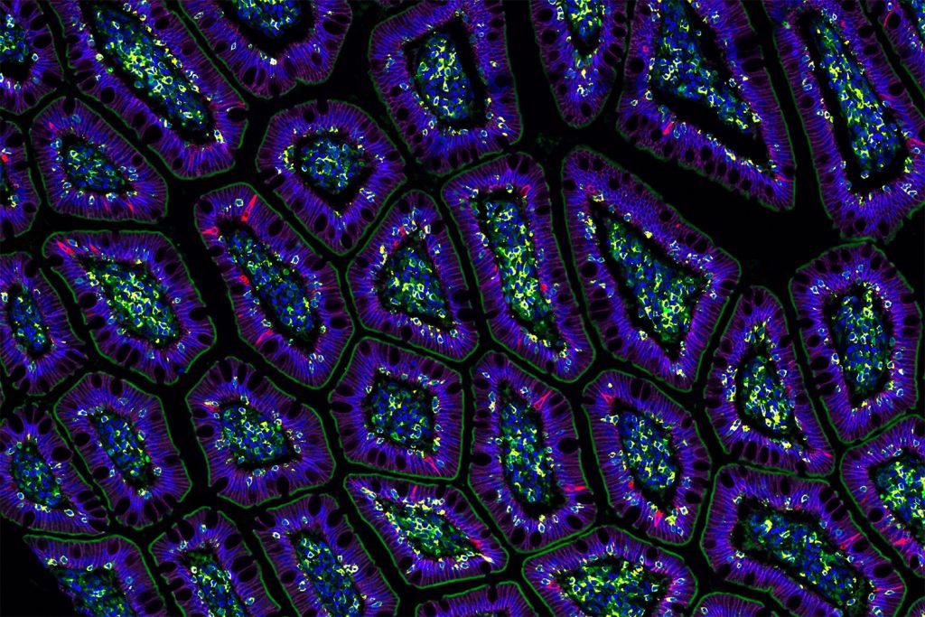
Knowing where a protein is expressed within a cell gives you clues to its function. If it’s in the mitochondria, it’s probably involved in energy metabolism. If it’s in the nucleus, it’s probably involved in gene regulation. I sometimes compare it to a house – if I know you’re in your kitchen or in the laundry room, I can make an educated guess about what you’re doing.
Proteins execute all of the functions in the human body. Most drugs are designed to target them. A protein needs to be in the right place to perform its function, because most protein functions happen through interactions. If a signaling protein ends up in the wrong compartment of a cell, for example, it might either break that signaling or give false signaling.
A single protein expressed in the wrong place can lead to cellular dysfunction and disease. There are known examples across many types of diseases, including cancer, immunological disorders, and neurological conditions. That’s why we have to measure proteins – because proteins and their spatial organization are the network that makes this complex system function. With better understanding of the protein architecture and wiring of a cell, we might be able to relocalize proteins therapeutically and restore cellular function.
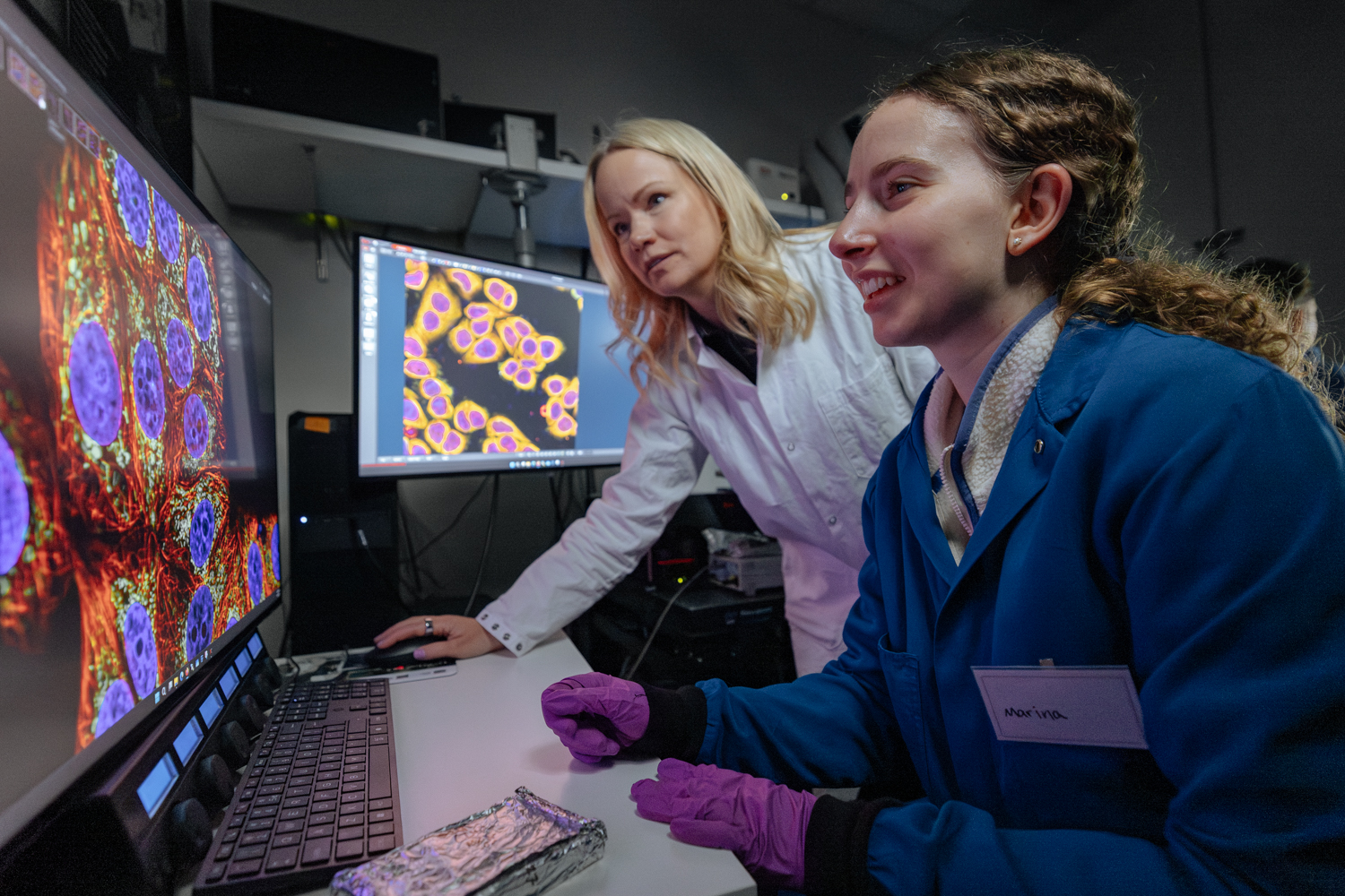
You can only visualize so many things at once with microscopy – the colors overlap and we can routinely image four colors at a time. So we can see a protein plus three markers to orient us in the cell. We’ve done this experiment tens of thousands of times, changing out the protein of interest, so we have many measurements of proteins with these reference landmarks, but they’re all separate.
We trained a model, called ProtiCelli, that essentially allows us to see all 13,000 proteins inside a single human cell at once. Now we can measure and analyze this massive image and harvest a lot more information. This is an example of how AI is breaking barriers and unlocking capabilities we’ve never had before.
“Now we have the tools to actually model from the data.”
Going forward, we want to do more than just build a map. If you really want to understand a cell as a system and model it, you need to perturb that system. If we remove a protein from the cell, relocalize a protein within the cell, or add a drug, what happens? That’s the next stage we’re thinking about right now: how we can move from baseline measurements to these highly informative perturbation measurements that form the foundation for building virtual cell models.
The ultimate dream, of course, is a virtual cell model that you could prime with someone’s genetic background and test how it would respond to a drug treatment. I think we’re far from that right now. But we’re building lab-in-the-loop systems that use these models to guide what experiments to do, so that instead of doing 10,000 experiments, we can do 1,000 highly informative ones, and then update the model. It’s changing the way we do biology.
I am a strong advocate for open science, making data available and useful for everyone, and accelerating science overall. That’s why I’ve chosen to be in academia. The Human Protein Atlas, a database that provides basic knowledge about protein localization and expression across human cells and tissues, is being used by millions of people all over the world to help identify targets for understanding diseases and developing better drugs. Resources like these are the foundation that allow other researchers to do their science faster.
Basic biology research to understand cells, these incredible complex systems that are the basis of all life, is not a trivial task. Most disease interventions and drugs that are developed come from basic biological discoveries. We have to start there.
This is a new chapter where we’re not just doing biology the way we’ve always done it, but deploying new technologies and automated lab-in-the-loop systems that we built here at Stanford. It’s a really exciting time.
For more information
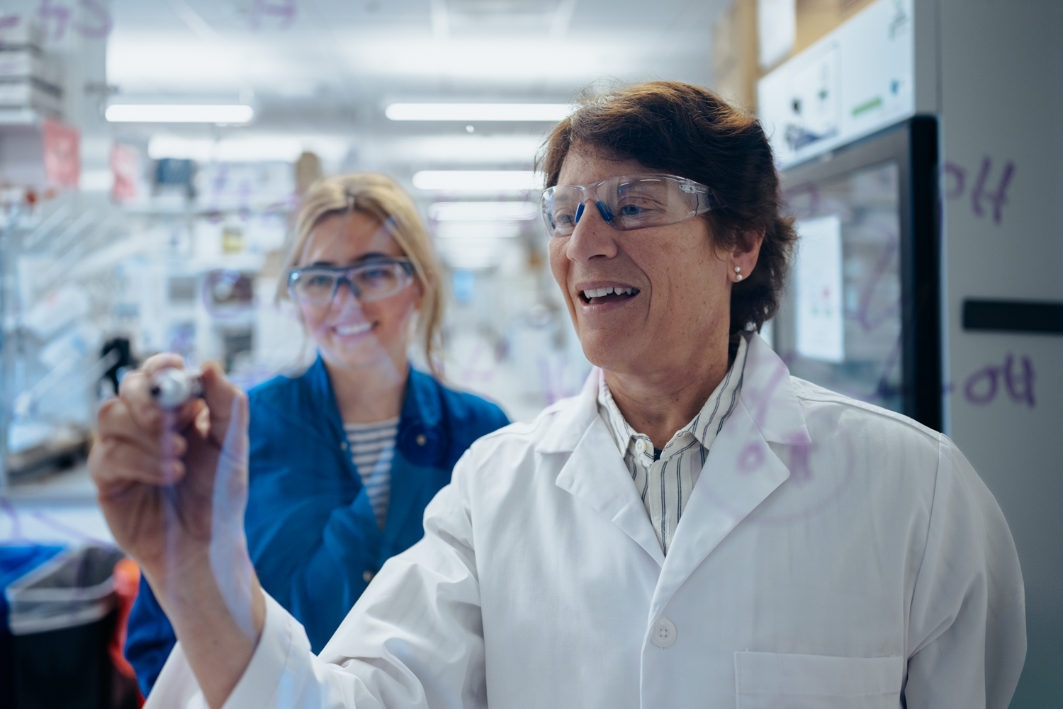
Emma Lundberg is an associate professor of bioengineering and of pathology in the schools of Engineering and Medicine. She is also a member of Bio-X, Wu Tsai Human Performance Alliance, and Wu Tsai Neurosciences Institute.
Photographer
Andrew Brodhead