In the “Research Matters” series, we visit labs across campus to hear directly from Stanford scientists about what they’re working on, how it could advance human health and well-being, and why universities are critical players in the nation’s innovation ecosystem. The following are the researchers’ own words, edited and condensed for clarity.
When I was a graduate student at Stanford, I cared for a young child newly diagnosed with diffuse intrinsic pontine glioma, or DIPG – a fatal brainstem tumor and the most common high-grade glioma in children. I was there when we told her family the prognosis: less than a 1% five-year survival rate. This was about 20 years ago, and at that point, there was nothing we could offer but palliative radiation. No chemotherapies worked. We didn’t even know what caused it.
These are previously healthy kids, often 6 to 8 years old, who come in with maybe a crossed eye and another symptom or two – and then the disease takes hold and progresses rapidly. They become unable to move, speak, or communicate, but remain fully aware.
Thinking back to that first patient, I get choked up. I remember thinking, “How is there no research on this disease?” And part of the reason was that there were no experimental models – no cell cultures, no mouse models. Because the tumor grows in the brainstem and diffusely infiltrates the nervous system, there was almost no tissue available for research at that time. It’s not a mass you can surgically remove and study; it’s like trying to get glitter out of Jell-O.
As a PhD student, I had learned to culture or grow hippocampal stem cells from early post-mortem brains, and I wondered if we could apply that to culturing DIPG cells in the postmortem period. I thought families might be willing to donate tissue, so we could understand where this cancer comes from and work to find ways to treat it.
“I’m filled with hope that this approach will ultimately lead to better outcomes for this otherwise lethal cancer.”
Studying neural precursor cells in pediatric autopsy samples, we found a unique population of oligodendrocyte precursor cells (OPCs) that increases in a part of the brainstem called the ventral pons around ages 6 to 8 – exactly when DIPG typically arises. These OPCs give rise to oligodendrocytes, which produce myelin, the insulating coating on neurons that speeds up signal transmission.
That convergence – between a developmental surge in OPCs in the ventral pons and DIPG’s typical onset window – offered the first real clue that this cancer might arise from early glial precursor cells.
It was a finding that set the course for the rest of my research.
At the same time, a new idea was emerging that nervous system activity might influence myelin, that myelin may have the ability to change in shape and efficiency throughout life – but this idea of “myelin plasticity” was very controversial in the field. And so I decided that I would take on that question, leveraging new techniques in modern neuroscience developed here at Stanford, like optogenetics, to ask these questions about brain cancers and glial cells.
We opened my lab in 2011, and the first experiments produced just jaw-dropping results. We found the first direct evidence for myelin plasticity, which helped reinvigorate the field. We’re still working to understand all the ways that this really fascinating, newly recognized dimension of neuroplasticity influences brain functions.
These normal versions of the glial precursor cells that DIPG appears to arise from connect to neurons for reasons we still don’t fully understand. That led us to ask: Could cancer cells be connecting in this way, too? And we discovered that yes, they are. There are electrophysiologically functional synapses between neurons and the cancer cells invading those circuits. This synaptic integration is fundamental to tumor growth, invasion, and progression.
With a cancer that is structurally and electrically integrating into the brain, we thought that one way to “dis-integrate” it would be with a cellular immunotherapy like CAR T-cell therapy. In collaboration with Stanford leaders in immunotherapy, we’ve now developed a CAR T-cell therapy for the fatal pediatric brainstem tumor DIPG. This is an intensive form of treatment in which a patient’s own immune cells are modified to attack cancer cells. It’s still in early-phase clinical trials, but over the past five years, we’ve already seen enormously encouraging results. Some patients have experienced dramatic tumor reductions and improvements in neurological function. One child even had a complete response that’s lasted four years, giving us real hope that we can achieve similar outcomes for more patients.
We’re still in the early stages of optimizing this therapy and figuring out how to deliver it most safely. But I’m filled with hope that this approach will ultimately lead to better outcomes for this otherwise lethal cancer.
None of this progress would have been possible without the NIH. Their support has been foundational to every stage of my career – from my PhD and postdoc to launching my lab and making key discoveries, including the identification of neuron-to-cancer synapses.
Our breakthroughs in brain cancer therapy grew directly from studying the fundamental biology of how things work – and how they go wrong.
For more information
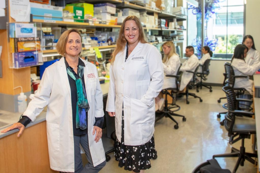
Michelle Monje is the Milan Gambhir Professor in Pediatric Neuro-Oncology and professor of pediatric neurology at Stanford Medicine, where she is also a professor, by courtesy, of neurosurgery, of pediatrics, of pathology, and of psychiatry and behavioral sciences.
She is a member Stanford Bio-X, the Institute for Stem Cell Biology and Regenerative Medicine, the Maternal & Child Health Research Institute, the Stanford Cancer Institute, and the Wu Tsai Neurosciences Institute.
Photographer
Andrew Brodhead