In brief
- Some immune cells undergo a process called NETosis, in which the cell swells like a balloon and then breaks open, releasing its DNA to trap invading pathogens.
- Better understanding the steps involved in NETosis could lead to new treatments for autoimmune diseases, cancer, and more.
- The team found that, while still stored in the nucleus as chromatin, DNA unwinds itself and releases proteins out of the nucleus into the cell. That flood of proteins triggers an influx of water into the cell, causing it to burst.
- This is the first example of DNA inside the nucleus nongenetically altering the mechanical properties of the entire cell.
Immune cells are the elite athletes of the human body. While other cells stay in place, immune cells travel throughout our bodies, squeezing through small channels, stretching themselves and sampling the surfaces of other cells, and swallowing up invaders. One of their most extreme behaviors is NETosis, a process in which certain immune cells burst and release their DNA as a way to trap pathogens.
NETosis, which is both critical when clearing infections and a liability in autoimmune diseases, involves several key steps: DNA, normally wound tightly into chromatin in the nucleus, unwinds itself; the nucleus breaks open to release DNA into the cell; the cell membrane ruptures, and the innards spill out. But the order of events and what triggers each step are poorly understood.
In a recent study published in Nature Communications, a team led by Hawa Racine Thiam, an assistant professor of bioengineering and institute scholar at Stanford University, discovered a key step in NETosis progression. They uncovered a previously unknown mechanism through which DNA, while stored inside the nucleus, puts stress on the cell membrane until it pops like an overfilled water balloon. The team also showed that they could leverage this mechanism to either prevent or speed up NETosis in cells. By better understanding how cells rupture and identifying the levers that control key steps, scientists can develop new therapies for autoimmune diseases, cancer, and more.
“Immune cells teach us what is possible,” said Thiam. “By studying extremes like NETosis, we are not just learning how immune cells behave, but we are also uncovering fundamental biology and learning what perhaps other cells are capable of. And if we understand those behaviors, we can start to engineer those behaviors.”
Unspinning a yarn
Manasi Sawant, a postdoctoral scholar and co-lead author of the paper, has been studying what happens to chromatin inside the nucleus during NETosis. “Imagine a tangled ball of yarn. The thread of yarn is DNA, and it is twisted and molded into a tight space,” said Sawant. “During some processes, like NETosis, the chromatin decompacts, which means the DNA loosens up.”
Chromatin also unwinds itself at other times, like when healthy cells replicate themselves or when cancer cells mutate and become drug resistant. By studying the steps by which chromatin unwinds during NETosis, Sawant hoped to learn more about these other processes, too.
One way to study the unwinding is to look for the spools, the proteins that bind to DNA yarn and help keep it wrapped up tightly. Two of these proteins are called H1 and H1Pɑ. The amount of these proteins floating freely in the nucleus, compared to the amount wrapped up in chromatin, gives scientists a measure of how much the chromatin has unwound itself.
Sawant’s experiments showed that as NETosis proceeds, H1Pɑ leaves the chromatin, as expected. But she could not find H1Pɑ floating around in the nucleus.
At the same time, Aidan Cabral, a bioengineering PhD student and a co-lead author of the study, was studying what leads to cell membrane rupture during NETosis. Other scientists had guessed that, once free from the nucleus, the unwound and wiggling strands of chromatin press on the membrane until it pops. But Cabral found that the membrane tension increased before the chromatin ever left the nucleus.
“This was really surprising, that changes in chromatin compaction in the nucleus can have a huge, long-range impact on the mechanics of the entire cell,” said Cabral.
The mystery remained: What was putting pressure on the cell membrane?
A grain of salt
Cabral and Sawant soon realized that the two phenomena they were studying were connected. They found that the release of HP1ɑ, H1, and other similar proteins from chromatin during NETosis correlates directly with an increase in pressure on the cell membrane and in cell volume until it eventually bursts open. All the while, the chromatin stays nestled inside the nucleus.
To understand how these proteins trigger membrane rupture, think back to grade school lessons in osmosis. Water will flow from a less salty environment into a saltier one until the salt concentration on both sides equalizes.
The team has proposed that H1, HP1ɑ, and the other spool-like proteins are a lot like those granules of salt. As the number of those free-floating protein molecules – both inside and outside the nucleus – increases, channels on the surface of the cell open to let water in until it pops.
To test their hypothesis, they changed the osmolarity, or effective “saltiness,” of the liquid around the cells. When the outside had a lower osmolarity than normal, water flowed inside the cell more quickly, leading the cell to burst. When the osmolarity of the outside was greater than that of the inside, water flowed out of the cell, decreasing the cell volume and preventing the cell from breaking apart.
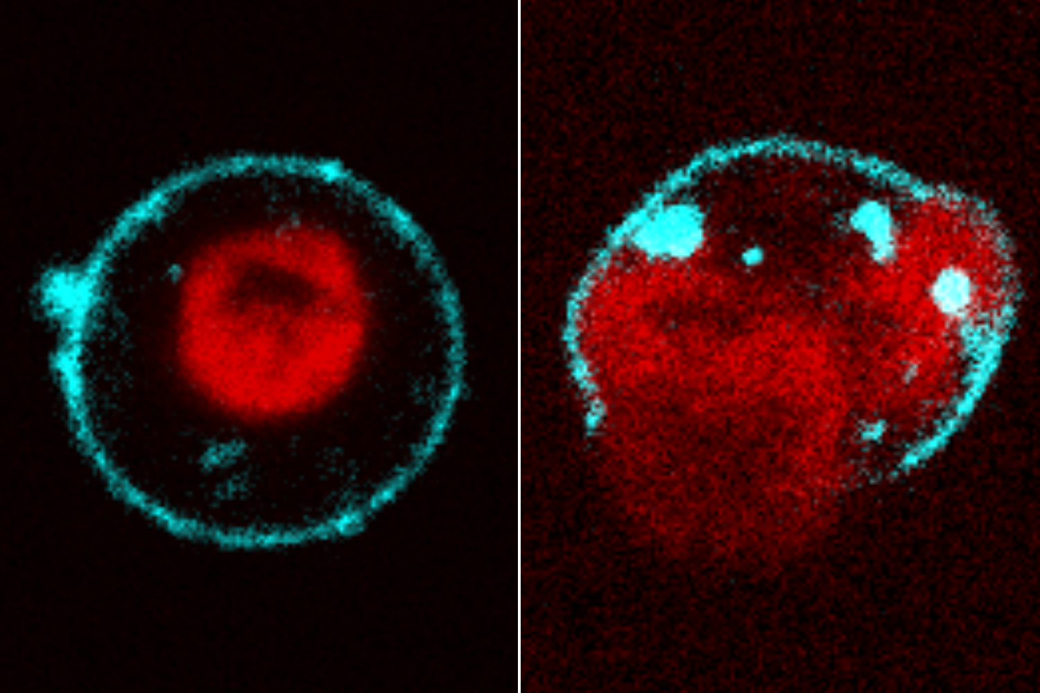
The authors showed that the osmolarity, or overall “saltiness,” of the cell leads to an influx of water into the cell, which in turn causes the cell membrane to rupture. When the environment outside the cell is less “salty” than normal (left), water flows into the cell more quickly and the cell ruptures faster. When the environment outside the cell is “saltier” than normal (right), the cell ruptures more slowly. | Minwoo Kang and Manasi Sawant
Beyond the blueprint
This mechanism represents a fundamentally new role for chromatin. DNA is the cell’s blueprint, and a particular gene’s placement in the chromatin is partly how the cell controls how much of that gene is expressed at a given time. “But here, changing how the DNA is packed changes the mechanics and the fate of the cell through physics, not genetics,” said Thiam.
This new understanding pointed the team to a way to prevent a cell from bursting. Membrane tension and cell volume are regulated by a series of tiny pores on the surface of the cell, called ion channels. The team showed that if they block some of these channels, they can stop a NETosing cell in its tracks, even after chromatin starts to decompact. This means that drugs targeting these channels could prevent overactive NETosis, potentially helping patients with autoimmune diseases.
Membrane tension and cell volume are regulated by a series of tiny pores on the surface of the cell, called ion channels. A cell that normally undergoes NETosis (left) will not rupture when treated with inhibitors of those ion channels (center, right), even after chromatin starts to decompact. | Manasi Sawant
They also put their newfound understanding to a bigger test, tricking a cell that normally does not undergo NETosis into inflating like a balloon. When they took a kind of bone cancer cell called a U2OS cell and added a molecule that causes the chromatin to decompact, the membrane tension increased, much like immune cells do during NETosis.
“While this is still a proof of concept, it shows that once you understand the process, you can start to imagine how you would engineer it: to make a cancer cell pop, to prevent overactive NETosis, or even to use immune cells as a way to package and deliver drugs to the site of infection,” said Thiam. “And we can imagine this because we study extremes.”
For more information
Thiam is also an assistant professor of microbiology and immunology and a member of Stanford Bio-X and of the Stanford Maternal & Child Health Research Institute (MCHRI). Other co-authors include postdoctoral scholar Minwoo Kang and Liangqi Xie of the Cleveland Clinic Lerner Research Institute.
The study was supported by a Stanford Graduate Fellowship, Stanford School of Medicine Dean’s Postdoctoral Fellowships, the Chan Zuckerberg Biohub San Francisco, the David and Lucile Packard Foundation, Stanford Bio-X, the Koret Foundation, and the Esther Ehrman Lazard Faculty Scholar Award.
Video and image adapted, with permission from the authors, from Cabral, A.T., Sawant, M., Kang, M. et al. “Chromatin decompaction within the nucleus increases plasma membrane tension promoting NETosis execution, independently of transcription,” Nature Communications (2026). https://doi.org/10.1038/s41467-026-76578-1
Media contact
Rebecca McClellan
Sarafan ChEM-H
rmcclell@stanford.edu
Writer
Rebecca McClellan