Inside a peculiar printer, acoustic pulses shoot through a blood sample of an ailing patient, pushing individual blood droplets onto a piece of paper. Among the tens of millions of droplets in the final print, only a few matter. Those are the ones that contain bacteria that could threaten the life of that patient.
This bio-printer is being developed by a Stanford University team that pulls together expertise from the schools of Engineering, Medicine, Humanities and Sciences and Business. Paired with an advanced way of scanning and identifying bacteria – which the group is also developing – this technology could speed diagnosis of bacterial blood infections and reduce the use of ineffective antibiotics.
A video (slowed 2500x) showing tests of the acoustic ejection technique that will be used in the printer. The researchers can eject these droplets from an open surface, which avoids contamination and clogging, problems that are common to bioprinting. (Video credit: Fareeha Safir)
Led by Jennifer Dionne, associate professor of materials science and engineering, the project is among the 2017 Stanford Catalyst for Collaborative Solutions grant recipients. All of the Catalyst awarded projects address complex societal challenges through innovative interdisciplinary solutions, including this project’s focus: How do we achieve effective yet affordable healthcare everywhere?
“This is a truly interdisciplinary project,” said Dionne. “Our team’s collaboration across the schools of Engineering, Medicine and Business has given rise to new ideas that may not have emerged in a traditional setting.”
Once the team has a full working prototype, they plan to focus on making it as low-cost as possible. With some adjustments, this system may also be able to test other bodily fluids, such as saliva or urine, and identify for other biological indicators of disease.
A microbial culture shift
The current gold standard for screening bacterial infections and deciding which antibiotic to administer involves growing blood-born bacteria in the lab, but, in some cases, that may take too long.
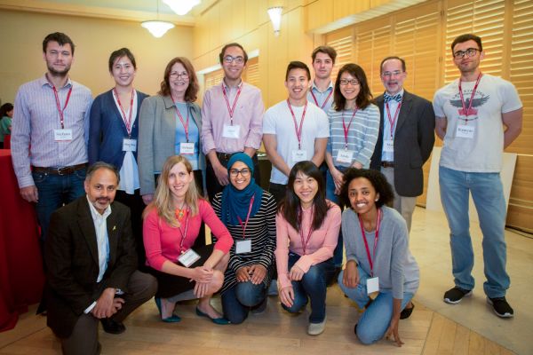
Some members of the Catalyst collaboration who are working on rapid screening of bacterial infections in the bloodstream (top row, left to right) Stefano Ermon, Shing-Shing Ho, Stefanie Jeffrey, Amr Saleh, Neal Jean, David Huland, Sindy Tang, Mark Holodniy, Kamyar Firouzi and (bottom row, left to right) Sam Gambhir, Jen Dionne, Fareeha Safir, Fengjiao Lyu, Loza Tadesse. (Image credit: Saul Bromberger)
“Patients with bacterial infections in their bloodstream may develop sepsis. If left unchecked, sepsis can progress to multi-organ failure fairly quickly, and unfortunately, some septic patients will die,” said Stefanie Jeffrey, John and Marva Warnock Professor in the Department of Surgery. “So, they are treated with a combination of strong antibiotics that cover a large spectrum of bacteria, hoping that these antibiotics will treat the particular bacteria that may be causing the infection.”
Although those powerful antibiotics often fight the infection, they don’t always work well or could be associated with toxic side effects. They could also increase the likelihood of bacteria becoming resistant to antibiotics.
The team designed the printer to be speedier than growing cells in the lab. The print out can be scanned with a laser and the colors of light that bounce off the page reveal each cell type present within the droplets. Machine-learning algorithms analyzing the light can detect the presence of bacteria, down to the specific strain and how it would respond to antibiotics. All it takes is a matter of minutes.
Once the system is complete, the entire blood droplet printing and scanning process will be automated, potentially at the point-of-care, reducing the work load of health care professionals and saving health care facilities time and money.
Accurate detection
In preliminary work, the researchers have tested their algorithm on 31 strains of bacteria grown in the lab – representing about 95 percent of bacterial infections seen at Stanford Hospital – and have shown their algorithm can recommend the correct treatment with over 98 percent accuracy, on average. Additional tests, looking at six classes of bacterial infections from actual patients, resulted in over 80 percent correct diagnoses for just a single cell in the patient sample, and over 90 percent accuracy when 10 bacterial cells were present. The researchers have also confirmed that cells survive the printing process.
In addition to beefing up the algorithm’s detection, the team is devising ways of making the signal brighter. They are working on metallic nanostructures that enhance the signal absorbed and produced by the laser light interacting with the cells.
“The nanoparticles efficiently absorb and scatter light,” explained postdoctoral scholar Amr Saleh. “The Rose Windows in Stanford Memorial Church get their color from a similar process.”
A major hurdle remains in testing whether their results from pure bacteria samples will hold up when analyzed in whole blood. Material in blood, like proteins and other cellular and extracellular components, could interfere with the signal from the bacteria, Dionne said.
Once the group proves the prototype system can quickly and accurately identify bacteria in whole blood samples, the researchers plan to reconfigure it into the most affordable version for use in low-resource areas, like rural clinics or places without access to cell culture facilities. This means recreating the functions of equipment that is worth tens of thousands of dollars with parts that together costs only tens of dollars. Meanwhile, they are also working with Robert Chess and Supriya Hobbs in the Graduate School of Business to evaluate the market for this technology and ensure what they develop meets market needs.
“Because of Stanford expertise in so many areas – nanoparticles, optics, droplets, acoustic printing, artificial intelligence and infectious disease – we can build on advanced technology and hopefully make it cost-effective, said Jeffrey. “We’re very fortunate that we’re able to bring together a collaborative team that covers many different areas to work toward one goal.”
Dionne is also a member of Stanford Bio-X, an affiliate of the Stanford Precourt Institute for Energy and a member of the Stanford Neurosciences Institute. Jeffrey is a member of Stanford Bio-X and a member of the Stanford Cancer Institute. Other collaborators on the work include Mark Holodniy, professor of medicine and a member of Stanford Bio-X, and Pierre Khuri-Yakub, professor of electrical engineering and a member of Stanford Bio-X, the Stanford Cancer Institute and the Stanford Neurosciences Institute.
Media Contacts
Taylor Kubota, Stanford News Service: (650) 724-7707; tkubota@stanford.edu
Author
Taylor Kubota