In brief
- Researchers used SLAC’s Linac Coherent Light Source to study the AspH enzyme – a potential therapeutic target for cancer treatment – and the isopenicillin N synthase (IPNS) enzyme, which is essential for creating all of the medicinally important penicillin antibiotics.
- By leveraging the power of ultrabright, ultrashort X-ray bursts, they found that these enzymes catalyze reactions in surprising ways.
- If scientists can better understand the structure and function of these enzymes, they could potentially use them as biomarkers and therapeutic targets for cancer, or for a method to develop new antibiotics.
Within biology, one group of enzymes, called 2-oxoglutarate-dependent oxygenases, is responsible for catalyzing a very wide range of reactions that play a role in everything from how genetic code is used by cells to synthesizing antibiotics.
Within the past several years, researchers have used ultrashort, powerful light pulses from X-ray free-electron lasers, including the Linac Coherent Light Source (LCLS) at the Department of Energy’s SLAC National Accelerator Laboratory, to better understand just how these enzymes catalyze reactions.
In two recent papers, researchers used LCLS to study the AspH enzyme – a potential therapeutic target for cancer treatment – and the isopenicillin N synthase (IPNS) enzyme, which is essential for creating penicillin. By examining those reactions step-by-step at mere quadrillionths of a second, they found that these enzymes activate reactions in surprising ways – leading to new insights that could inspire novel therapeutics and drug development.
“We have gone from examining static structures to making ‘molecular movies’ of the dynamics of biological systems,” said Roberto Alonso-Mori, senior scientist at SLAC. “It has really given us an understanding of the function of proteins.”
The research was led by Patrick Rabe and Christopher Schofield, research group leaders at the University of Oxford, U.K., in close collaboration with Jan Kern, senior scientist at DOE’s Lawrence Berkeley National Laboratory (Berkeley Lab). The research was published in Nature Communications and Nature Catalysis.
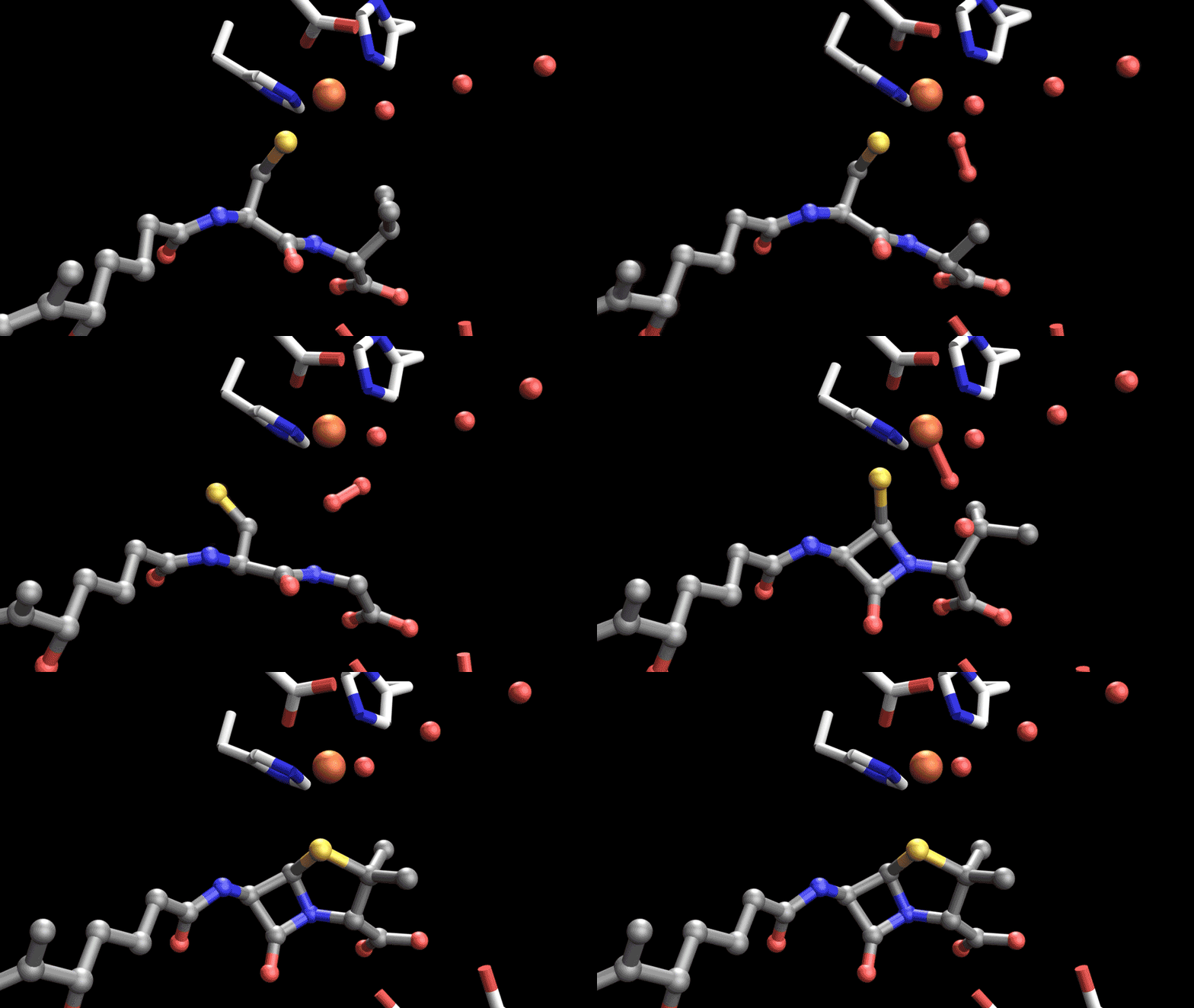
Animated molecular movie of penicillin biosynthesis: Each frame shows a different stage of the reaction as IPNS converts its substrate into penicillin. The animation was reconstructed from several time-resolved X-ray crystal structures collected at different reaction times, allowing the complete catalytic process to be visualized at atomic resolution. | Patrick Rabe / University of Oxford and Greg Stewart / SLAC National Accelerator Laboratory
Using X-ray free-electron lasers to identify potential pathways for therapeutics
In humans, the AspH enzyme is encoded by the AspH gene. Scientists have found that levels of AspH are increased in multiple cancers, especially the hard-to-treat brain cancer called glioblastoma. By examining the structure and function of AspH and how it is regulated, scientists could potentially use the enzyme as both a biomarker and a therapeutic target for cancer treatment.
Penicillin has a remarkable structure made up of two reactive rings, but as a consequence, it cannot be made by chemical synthesis. Instead, it is made by fermentation in microbial cells. During the process of manufacturing penicillin in cells, the IPNS enzyme catalyzes a vital early-stage reaction. Analyzing just how this reaction works could help scientists find a way to create synthetic modified penicillin that could overcome antibiotic resistance to better treat bacterial infections.
Researchers have studied how these enzymes catalyze reactions for years, but capturing intermediates – the molecules produced and then consumed in the middle of a reaction – has been challenging.
At LCLS, researchers use macromolecular crystallography with femtosecond pulses to observe what exactly happens during these reactions. Leveraging extremely bright and short X-ray bursts, they can do so in atomic detail, on timescales of millionths of a billionth of a second, and at room temperature without damaging the biological samples.
At the Macromolecular Femtosecond Crystallography (MFX) instrument, researchers create microcrystal samples of the enzymes and run them through a system that injects oxygen to induce the reaction. They then examine how X-rays from LCLS scatter off the sample in real time, turning those data into molecular movies of the reactions. “We have been working for many years to develop experimental protocols that allow us to precisely start the reaction in these fascinating enzymes and watch them perform complicated chemistry step by step in real time,” Kern said.
“We wanted to understand specifically how enzymes are controlling the intermediates,” Rabe said.
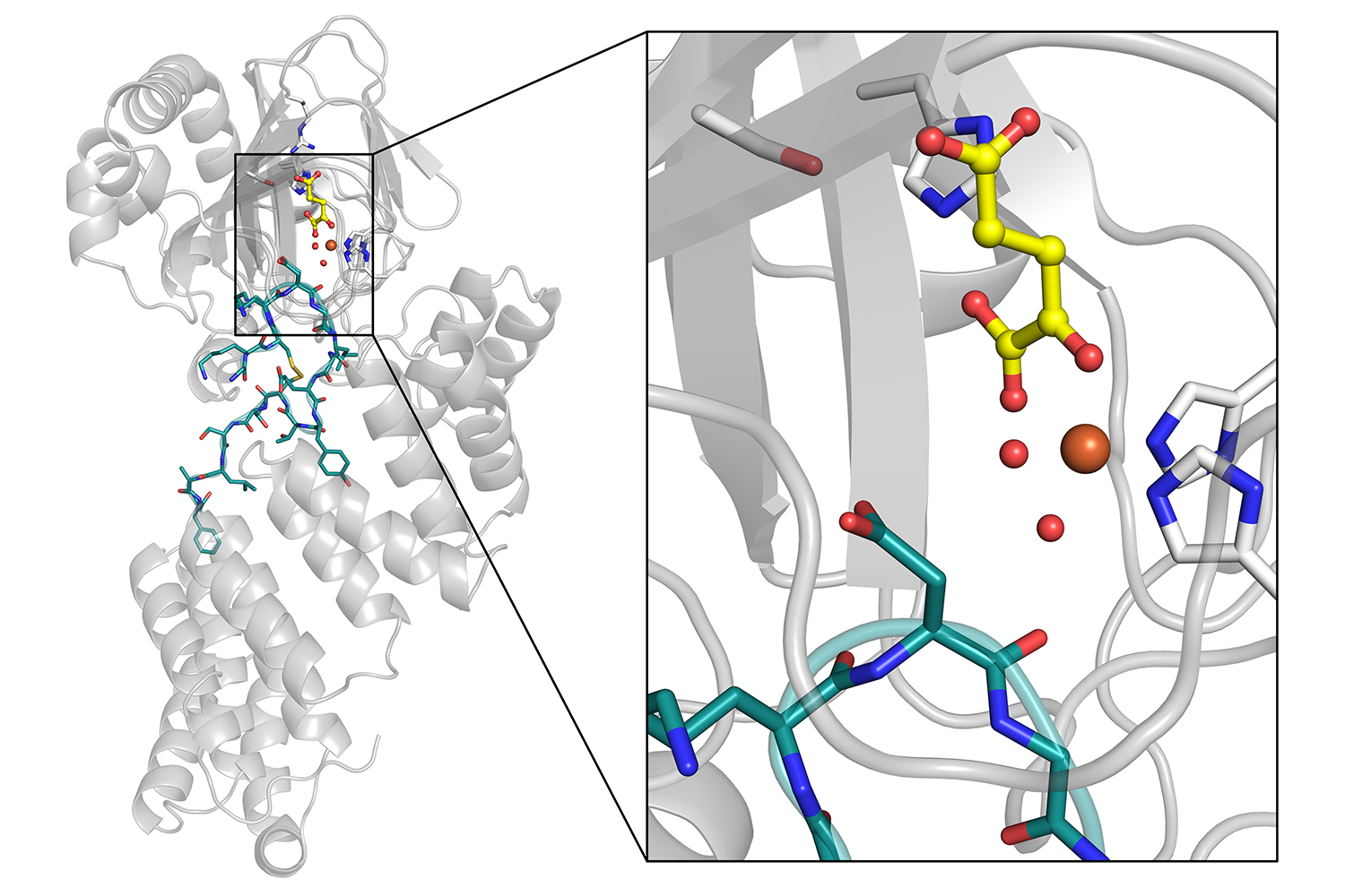
Structure of the human enzyme AspH: The enlarged view shows the enzyme’s iron-containing active site (orange sphere) bound to its protein substrate. Using time resolved X-ray crystallography, the researchers observed how AspH activates oxygen and hydroxylates proteins, providing new insight into a family of enzymes involved in oxygen sensing, development and cancer. | Patrick Rabe / University of Oxford and Greg Stewart / SLAC National Accelerator Laboratory
‘Molecular movies’ reveal surprising results
For both enzymes, researchers knew that iron played a key role in the reactions – but were surprised by how the steps played out. “Our observations from these experiments were not predicted by sophisticated calculations,” Schofield said.
For AspH, the researchers saw how the enzyme binds to a substrate and activates oxygen during catalysis. In particular, they saw that an iron-bound water molecule remains stable throughout the reaction, a factor that appeared to be important for catalysis. The research team knew that iron-bound water was essential for catalysis, but seeing this stability – and showing that if it’s disrupted, the reaction won’t proceed – could help scientists develop a pathway to treating AspH activity in cancer cells.
They also found that AspH only uses two metal-coordinating residues – amino acids that bind to specific sites in proteins – instead of three residues usually seen in related proteins. Scientists could ultimately use this information to engineer new proteins that could modify peptides for therapeutic benefit.
In the case of IPNS, the researchers took snapshots of the intermediates during the synthesis of the penicillin rings. They found that large parts of the enzyme are involved in this reaction – not just the iron-containing site with the substrate – and that there are key water molecules involved. In fact, the water molecules are used to control the outcome of the reaction.
“That was very surprising to us,” Schofield said. “Both that regions away from the active site are important and that these active site water molecules were crucial to catalysis.” This step-by-step framework of IPNS catalysis could help scientists engineer enzymes to make penicillin more efficiently or target disease in new ways.
A crucial next step is to understand the intricate involvement of water in catalysis. The team also wants to create compounds that either promote or inhibit the activity of these enzymes – as a potential cancer treatment, for example – to understand them even more.
International, multidisciplinary collaboration drives innovation
The team has worked together for years on these projects, with Rabe and Schofield traveling from the United Kingdom to work with Alonso-Mori at SLAC and Kern at Berkeley Lab. The team also includes researchers across disciplines who provide the wide range of expertise needed to develop and use the technology necessary to understand nature at such a small scale and work toward new therapies for hard-to-treat diseases.
“It’s a really nice consortium of biologists, chemists, physicists, computational analysts, and engineers that all come together to study this, because we all need each other,” Rabe said.
“Many of us dreamt about having experiments like this decades ago, so, to have these results and to be able to move forward with this research is a dream come true,” Schofield said.
For more information
The research was in part funded by DOE’s Office of Science and the National Institutes of Health. In addition to researchers from SLAC, Berkeley Lab, and the University of Oxford, the research collaborations included scientists from the U.S., U.K., Republic of Korea, Japan and Latvia. X-ray data were collected at LCLS, a DOE Office of Science user facility; PAL-XFEL, Republic of Korea; SACLA, Japan; and Diamond Light Source, U.K.
Citation: De Munnik et al., Nature Communications, 25 February 2026 (10.1038/s41467-026-69425-w); Rabe et al., Nature Catalysis, 09 October 2026 (10.1038/s41929-026-01618-4.
This story was originally published by SLAC National Accelerator Laboratory.
Writer
Emily Ayshford