In brief
- A study of the long-held motor homunculus model reveals a more complex organization of the motor cortex that affects body movement control.
- Research shows areas in the motor cortex encode information for multiple body parts, suggesting broader relationships than previously understood.
- The findings could enhance brain-computer interface development for paralysis patients, enabling more versatile control with fewer implants.
For almost a century, budding neuroscientists have been taught that the headband-like strip of brain tissue over our ears that controls our movements, called the motor cortex, contains an orderly map of our bodies. Brain cells concerned with moving each body part – from the tips of our toes to the tips of our fingers – are all laid out in sequence, as well as a large zone dedicated to our fabulously expressive faces and speech muscles. That’s the textbook account, anyway.
But that extremely satisfying model, known as the motor homunculus, doesn’t tell the full story, according to a new study by Wu Tsai Neurosciences Institute researchers, published June 17, 2026, in the journal Nature. The new study finds that many parts of the motor cortex have information about the entire body. The region that is predominantly associated with movement of the arms, for example, also has some information about movements of the face, and the face region has some information about movement of the legs.
“It was quite exciting to see these whole-body representations appear,” said Darrel Deo, an instructor in the Department of Neurosurgery at Stanford Medicine and the new paper’s first author.
Apart from a deeper understanding of the motor cortex, the results could also lead to better brain-computer interfaces (BCIs) – systems that translate brain activity into signals that control or interact with devices that aid people who’ve lost the ability to move or communicate because of neurological disease or paralysis. The new study suggests a single implant could provide access to a wider repertoire of movement-related control signals than previously appreciated, Deo said, rather than one implant for each affected region of their body.
Deo began the work as a Wu Tsai Neuro postdoctoral scholar in the lab of the late Krishna Shenoy and Jaimie Henderson, the John and Jene Blue - Robert and Ruth Halperin Professor and a professor of neurosurgery at Stanford Medicine. As part of that effort, Deo and lab co-director Frank Willett, an assistant professor of neurosurgery at Stanford Medicine, wanted to update the motor homunculus map with modern tools. “We were keen to revisit the motor cortex at a much finer scale,” Deo said.
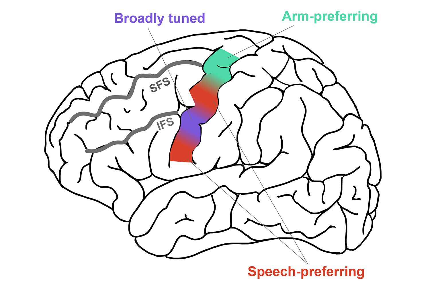
A map of the motor cortex highlighting regions tuned more for speech-related movements (red) and arm movements (blue-green) and a particularly broadly tuned region (purple). | Courtesy Darrel Deo
To do so, Deo and his colleagues studied eight participants who were already enrolled in ongoing BCI clinical trials – six from the BrainGate2 clinical trial and two from a separate clinical trial run through the University of Pittsburgh and University of Chicago. These participants were all implanted with microelectrode arrays capable of recording electrical signals from individual neurons in various locations along their motor cortex – something that wasn’t possible when the motor homunculus idea was first developed.
Then, Deo asked the participants to perform 45 different body movements, such as lifting a foot or turning a hand (or to attempt the movement if they were no longer able to move that body part) as he recorded activity from motor cortex neurons.
In each and every region the researchers looked at, Deo said, “there was distinguishable information about movements that span the entire body, not just the canonical body part the motor homunculus model would have suggested.” In other words, while one region might be most strongly activated for hand movements, it also contains signals concerning movements in the face, legs, and everywhere else.
The researchers also propose a new, modified map of the motor cortex that differs from the original homunculus model. In this new map, two areas tuned for speech flank the superior ventral precentral gyrus, which the team found was broadly tuned to all parts of the body. “It’s like a jack-of-all-trades area,” Deo said.
Those results could be a boon for BCI design. For example, a single electrode array implanted in the superior ventral precentral gyrus could be used to control robotic arms, communication devices, and more.
More generally, the study suggests it’s time to move beyond the motor homunculus’s strict partitioning of the motor cortex. “It’s a bit more intermixed than we thought,” Deo said.
For more information
This story was originally published by Wu Tsai Neurosciences Institute.
Writer
Nathan Collins