In brief
- Stanford Medicine researchers created CANVAS, an AI platform that infers cellular neighborhoods from routine pathology slides used for over a century.
- Trained on hundreds of lung cancer patients, CANVAS mapped 10 distinct cellular neighborhoods by decoding how cancer, immune, and stromal cells communicate.
- CANVAS predicts immunotherapy resistance more accurately than current clinical methods, potentially guiding treatment decisions across nine cancer types.
Understanding how cells within and around a tumor interact provides key information about a cancer’s architecture, a patient’s immune response to the disease, and even how susceptible the cancer may be to various types of treatment. But deducing these cellular “neighborhoods” using traditional techniques is time-consuming and expensive.
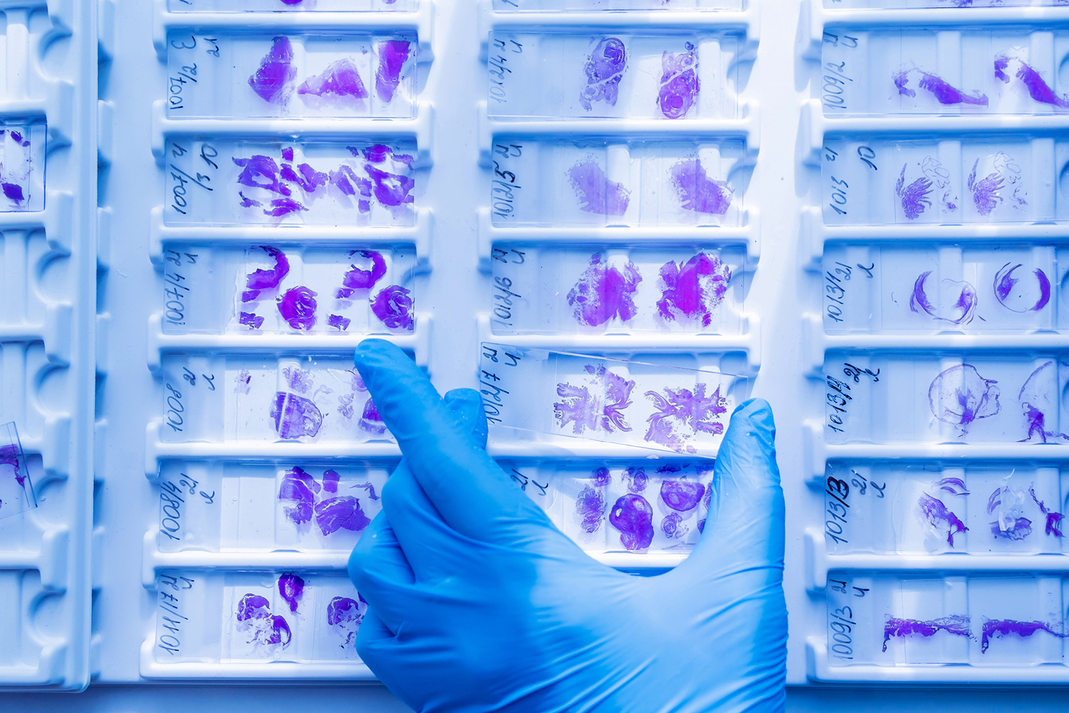
Now, Stanford Medicine researchers have developed an artificial intelligence platform that can predict the neighborhoods from microscopic slides containing slices of human tumor tissue – transforming flat pink and purple “pages” of limited information into riotously colored pop-up books chock-full of intel about the dynamic relationships and conversations between cells in space.
Using the platform, the researchers identified 10 discrete cellular neighborhoods – defined by interactions and molecular conversations among cancer cells, immune cells and stromal cells – in non-small cell lung cancer.
The microscopic slides, known as H&E slides for the two stains used to highlight the cells and extracellular structures the slices contain, have been the gold standard for disease diagnosis for more than a century. Essentially, every cancer patient who has undergone a biopsy or surgical removal of their cancer will have H&E slides of their tumor tissue.
The ability to infer complex relationships and cellular neighborhoods from existing slides, and correlate them to known patient outcomes, gives researchers a powerful new learning tool in the fight against many cancer types. For example, the researchers found that one cellular neighborhood, rich with a type of immune cell called neutrophils, correlates in lung cancer with a poorer overall prognosis and resistance to immunotherapy.
The accomplishment is possible thanks to a technique called CODEX developed in the Stanford Medicine laboratory of pathology professor Garry Nolan, PhD, the Rachford and Carlota A. Harris Professor. CODEX enables the detection of dozens of proteins and cell types within a tumor – yielding an unprecedented spatial map of relationships and cellular activity. But CODEX experiments can be slow and expensive.
“Understanding how cells interact within a tumor is particularly important because a tumor is a very complex ecosystem,” said Ruijiang Li, PhD, associate professor of radiation oncology. “But time and money limit the number of samples that can be analyzed with experimental techniques. We wanted to develop a tool capable of inferring this spatial complexity from existing resources like pathology slides.”
Li is the senior author of the research, which was published June 16 in Cell. Postdoctoral scholar Yuchen Li and research scientist Zhi Li are the lead authors of the study.
Ruijiang Li and his colleagues built an atlas of more than 18 million cells from 457 patients with non-small cell lung cancer and overlaid the CODEX results from these samples onto the microscope slides, matching them cell by cell to help the AI learn patterns on the slides that corresponded to the cellular neighborhoods predicted by the CODEX analysis.
They benefited from a previous AI tool called MUSK developed in Li’s lab that had been trained on 50 million medical images of standard pathology slides and more than 1 billion pathology-related texts.
They named the new AI platform CANVAS, for cellular architecture and neighborhood-informed virtual AI-driven spatial profiling.
“MUSK is particularly good at recognizing morphology patterns from slides,” Li said. “CANVAS uses those patterns to infer cellular neighborhoods – multicellular patterns in a complex tumor ecosystem.”
Researchers built an atlas of more than 18 million cells from 457 patients.
The cellular neighborhoods identified by CANVAS are defined not just by the prevalence and locations of cancerous and noncancerous cells in the sample, but also by their activities – which proteins they are making and what signals they are sending to the cells around them.
Some of the cellular neighborhoods, or habitats, showed signs of being immunosuppressive, tamping down an immune system trying to eradicate cancer cells. Others were found in the core of the tumor, or with clusters of immune cells or blood vessels.
After training CANVAS on the initial set of samples, Li and his colleagues then fed the platform images of archived slides from over 5,000 patients with nine cancer types to confirm the presence of 10 cellular neighborhoods and correlated their presence or absence with patient prognosis and responsiveness to immunotherapy.
They found that one cellular neighborhood, rich in neutrophils expressing proteins that may facilitate metastasis, was associated with worse outcomes for patients and a reduced responsiveness to immunotherapy drugs such as anti-PD-1.
The presence of this cellular neighborhood predicted responses to immunotherapy more accurately than approaches used now to determine if a patient is a candidate for the targeted treatments, suggesting that CANVAS could be a valuable tool to guide clinical decision-making.
“Next, we would like to validate CANVAS in clinical trials to confirm that we can predict patient prognoses and responses to specific treatment approaches,” Li said.
For more information
Researchers from the Broad Institute, Harvard Medical School and the MD Anderson Cancer Center contributed to the work.
The study was funded by the National Institutes of Health (grants R01CA290715, R01CA269599 and R01CA285456).
This story was originally published by Stanford Medicine.
Media contact:
Krista Conger: 650-725-5371; kristac@stanford.edu
Writer
Krista Conger