Unlocking pathways to break down problem proteins presents new treatment opportunities
Stanford researchers who previously pioneered a new kind of protein degradation have mapped out how the process works, which could lead to new drug therapies for diseases like Alzheimer’s and cancer, and for rare childhood enzyme disorders.

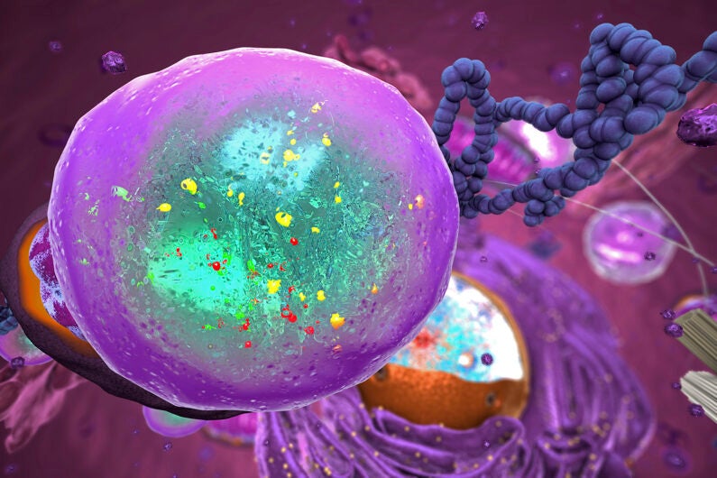
An illustration of a lysosome, which is cellular machinery that degrades proteins. Through studying precisely how a new class of molecule sends proteins to lysosomes – and what disrupts that process – researchers have unveiled possibilities for new drug therapies and medical treatments. (Image credit: Getty Images)
When targeting problem proteins involved in causing or spreading disease, a drug will often clog up a protein’s active site so it can’t function and wreak havoc. New strategies for dealing with these proteins can send these proteins to different types of cellular protein degradation machinery such as a cell’s lysosomes, which act like a protein wood chipper.
In a new study published in Science on Oct. 20, Stanford chemists have uncovered how one of the pathways leading to this protein “wood chipper” works. In doing so, they have opened the door to new therapeutics for age-related disorders, autoimmune diseases, and treatment-resistant cancers. These findings may also improve therapeutics for lysosomal storage disorders, which are rare but often serious conditions mostly affecting babies and children.
“Understanding exactly how proteins are shuttled to lysosomes to be broken down can help us harness the innate power of a cell to get rid of proteins that cause the human body so much harm,” said Carolyn Bertozzi, the Anne T. and Robert M. Bass Professor in the School of Humanities and Sciences and Baker Family Director of Sarafan ChEM-H. “The work done here is a clear look into a typically opaque intracellular process, and it’s shining a light on a new world of possible drug discovery.”
“The ability to understand the biology of this process means we can use inherent biology that already exists, and harness it to treat disease,” said Steven Banik, assistant professor of chemistry in the School of Humanities and Sciences. “These insights offer a unique window into a new type of biology that we haven’t really understood before.”
Stopping proteins from going rogue
While proteins often do a body good, like help us digest our food or repair torn muscles, they can also be destructive. In cancer, for example, proteins can either become part of the tumor and/or allow for its unchecked growth, cause devastating diseases like Alzheimer’s, and build up in the heart to affect how it pumps blood to the rest of the body.
To stop rogue proteins, drugs can be deployed to block a protein’s active site and thus stop it from interacting with a cell, which was the standard of therapeutic research for decades. Then 20 years ago, proteolysis targeting chimeras (PROTACs) burst onto the scene, which can engage bad-acting proteins that are already inside a cell, and send them off to be broken down in the lysosome.
PROTACs are currently in clinical trials and have shown efficacy in treating cancer. But they can only target a protein if it is inside the cell, which is only 60% of the time. In 2020, Stanford ChEM-H researchers pioneered a way to reach the other 40% of those proteins through lysosome targeting chimeras (LYTACs), which can identify and mark proteins that are hanging out around the cell, or on a cell’s membrane, for destruction.
These findings kicked off a new class of research and therapeutics, but exactly how the process worked wasn’t clear. Researchers also noticed that it was difficult to predict when LYTACs would be highly successful or fail to perform as anticipated.
New therapeutic targets
In this work, Green Ahn, then a Stanford graduate student and now a postdoctoral fellow at the University of Washington Institute for Protein Design, and lead author on the study, used a genetic CRISPR screen to identify and characterize the cellular components that modulate how LYTACs degrade proteins. Through this screening, the team identified a link between the level of neddylated cullin 3 (CUL3) – a protein that plays a housekeeping role in breaking down cellular proteins – and LYTAC efficacy. The exact tie isn’t clear yet, but the more neddylated CUL3 present, the more effective LYTACs were.
Measuring the level of neddylated CUL3 could be a test given to determine which patients are more likely to respond to LYTAC therapy. This was a surprise finding, said Bertozzi, as no previous research pointed to this correlation before.
They also identified proteins that block LYTACs from doing their job. LYTACs work by binding to certain receptors on the outside of the cell, which they use to shuttle bad proteins into lysosomes for degradation. However, the researchers saw that proteins bearing mannose 6-phosphates (M6Ps), sugars that decorate proteins destined for lysosomes, will take a seat on those receptors, meaning LYTACs have nowhere to bind. By throwing a wrench into M6P biosynthesis, an increased fraction of unoccupied receptors resulted on the cell surface, which could be hijacked by LYTACs.
New biology, new pathways for treatment of disease
In addition to helping develop LYTACs into more effective therapeutics, these discoveries could also lead to new and more effective treatments for lysosome shortage disorders – genetic conditions where the body doesn’t have enough or the right enzymes in lysosomes for them to work properly. This can cause toxic build ups of fat, sugars, and other harmful substances, which can lead to heart, brain, skin, and skeletal damage. One common treatment is enzyme replacement therapy, which utilizes similar pathways as LYTACs to travel to lysosomes where they can operate. Understanding how and why LYTACs work means that these enzymes could be delivered more effectively.
The researchers likened this work to an important discovery of how exactly the drug thalidomide works. It was originally prescribed in the 1950s for morning sickness to pregnant women, mostly in the United Kingdom, but was taken off the market in 1961 when it was linked to severe birth defects. However, in the 1990s, it was found to be an effective treatment for multiple myeloma. In 2010, researchers understood how: through degrading proteins, an observation which contributed substantially to the growing field of PROTAC research.
“LYTAC evolution is where the story of thalidomide and PROTACs was 15 years ago,” Bertozzi said. “We’re learning human biology that wasn’t known before.”
Other Stanford co-authors include former postdoctoral fellows Nicholas Riley and Roarke Kamber, PhD candidate Salvador Moncayo von Hase, and Michael Bassik, associate professor of genetics in the School of Medicine. Simon Wisnovsky of the University of British Columbia is also a co-author. Banik is an institute scholar at Sarafan ChEM-H and a member of the Wu Tsai Neurosciences Institute and Stanford Bio-X. Bassik is a member of Bio-X, the Stanford Cancer Institute, the Wu Tsai Neurosciences Institute, and Sarafan ChEM-H. Bertozzi is a member of the Wu Tsai Neurosciences Institute, Bio-X, the Stanford Cancer Institute, and the Maternal & Child Health Research Institute (MCHRI), as well as an investigator at the Howard Hughes Medical Institute.
This work was funded by the National Institutes of Health, the National Science Foundation, the Stanford Center for Molecular Analysis and Design, the Canadian Institutes of Health Research, the Natural Sciences and Engineering Research Council of Canada, the Cancer Research Society, and the Canadian Glycomics Network, and the Burroughs Wellcome Fund. Bertozzi is the founder of Lycia Therapeutics, where Banik is also on the Scientific Advisory board.
