
Extreme cold could help Stanford researchers reveal herpesvirus infection
Researchers don’t know much about how viruses like those that cause chicken pox infect cells. A super-cold form of electron microscopy could change that, potentially paving the way for new treatments and vaccines.
The funny thing about the virus that causes chicken pox is that no one knows for sure how it or many of its herpesvirus cousins invade and infect cells. It’s a bit of a problem: Without that knowledge, it’s been hard to find better ways to treat and prevent not just chicken pox, but other diseases caused by closely related viruses, like cytomegalovirus, Epstein-Barr virus and shingles – a painful condition related to chicken pox.
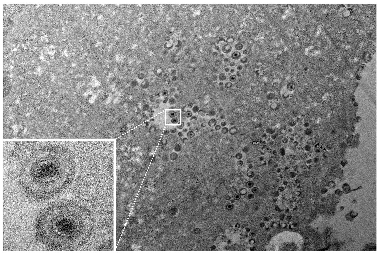
A transmission electron microscope image shows the varicella-zoster virus. An advanced version, cryogenic electron microscopy, could reveal even more detail, including how the virus infects cells. (Image credit: Stefan Oliver)
Now, Stanford virologists are working with scientists at the new Stanford-SLAC Cryo-Electron Microscopy facility to take a new look at how herpesviruses infect cells. With support from a Stanford Bio-X seed grant, they are taking some of the most detailed pictures ever of proteins on the surface of the chicken pox virus, also known as varicella-zoster virus. These images may soon reveal clues about how to block herpesvirus infections, said Stefan Oliver, a senior research scientist in the lab of Ann Arvin, the Lucile Salter Packard Professor of Pediatrics and a professor of microbiology and immunology.
“We’re using cryo-EM technology to look at the bigger picture quite literally,” Oliver said.
The key to the new images, Oliver said, is a relatively new technology called cryogenic electron microscopy, or cryo-EM. In the past, if researchers wanted to study how a virus uses proteins on its surface to infect cells, they would first produce shortened forms of virus proteins and crystalize them. By scattering X-rays off that crystal, teams could infer the structure of the protein, which could yield information about how it works.
The problem, Arvin said, is that the crystalized form doesn’t necessarily have the same shape as a protein as it exists on a virus or when it is made in infected cells. What’s more, X-ray crystallography can’t reveal how the shapes of those proteins change as they pry their way into cells, because it only captures the protein in one state.
Cryo-EM solves those problems, said Arvin and Oliver’s collaborator Wah Chiu, a professor of photon science, of bioengineering and of microbiology and immunology. By flash-freezing viruses at temperatures hundreds of degrees below zero, essentially stopping their motion while still preserving their structure, researchers can use electron microscopes to take pictures of viruses and their proteins. It’s a data intensive process – the cryo-EM lab works closely with the High Performance Research Computing Center at SLAC – but the end result is images with atomic-level detail, something that isn’t possible any other way.
So far, Oliver, Arvin and Chiu have begun to gather images of the varicella-zoster virus and a protein that helps the virus get into cells, glycoprotein B, along with an antibody to the protein. Those pictures revealed where the antibody binds to the protein, information that could help other researchers design molecules to interfere with infection. The next step, Chiu said, is to take pictures of the virus alongside actual cells, which would allow the team to see the varicella-zoster virus and its proteins at various stages of infection.
With that information in hand, Arvin said, “we’ve got a much better idea of how to interfere with the process of infection.” Down the line, that knowledge could lead to new ways to prevent chicken pox in children who can’t get the standard vaccine and better treatments for shingles and its aftereffects. It could also set the stage for a better understanding of other herpesvirus infections, Arvin said.
Arvin is a member of Stanford Bio-X, the Maternal & Child Health Research Institute, the Stanford Cancer Institute and the Wu Tsai Neurosciences Institute. Chiu is a member of Bio-X, Stanford ChEM-H, and the Wu Tsai Neurosciences Institute.
To read all stories about Stanford science, subscribe to the biweekly Stanford Science Digest.