October 3, 2019
Stanford chemist develop ‘infrared vision’ for cancer immunotherapy
A new technique employs a bright infrared light that can pass through millimeters of tissue to illuminate tumors deep inside the body.
See video here.
By Ker Than
Stanford chemists have developed a new deep-tissue imaging technique that can see beneath the skin of living subjects to illuminate buried tumors with unparalleled clarity.

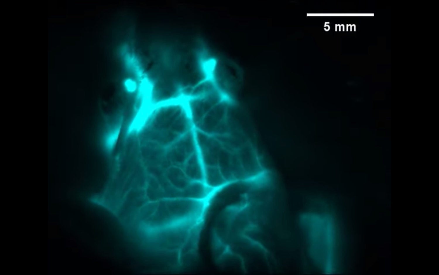
The brain vessels of a live mouse are illuminated in infrared light by erbium nanoparticle probes. (Image credit: Yeteng Zhong and Hongjie Dai)
In a new study published in the September 30 issue of the journal Nature Biotechnology, the researchers demonstrate how their technique can be used to predict the response of cancer patients to immunotherapy and to track their progress following treatment.
“We call this infrared vision for non-invasively peering into biological tissues,” said study leader Hongjie Dai, the J.G. Jackson and C.J. Wood Professor in Chemistry in Stanford’s School of Humanities and Sciences.
The technique relies on nanoparticles containing the element erbium, which belongs to a class of so-called rare-earth minerals prized by chemists for their unique ability to glow in the infrared.
The team covered the nanoparticles in a chemically engineered coating that helps the particles dissolve in the bloodstream and also makes them less toxic and exit the body quicker. In addition, the coating provides anchoring points for molecules that that act like guided missiles to locate and attach to specific proteins on cells.
Clearing the fog
Under a low-powered LED light, the erbium particles bask targeted tissues or even individual cells in a bright infrared glow that can be seen deeper and with finer resolution than conventional imaging techniques, the researchers say. “It is like seeing while driving on a highway before and after the fog evaporates on a winter day,” said study co-first author Yeteng Zhong, a postdoctoral researcher in Dai’s lab. “The combined imaging depth, molecular specificity and multiplicity, and spatial and temporal resolution are unattainable by previous techniques.”
In a video highlighting the power of their nanoprobes, the branching blood vessels of a brain inside a living mouse are limned in a teal light and shift as the creature moves its head. “Our approach allows for seeing into an intact mouse brain while conventional approaches see only the scalp,” said study co-first author Zhuoran Ma, a graduate student in Dai’s lab.
In the study, the researchers show their technique can identify tumors in mice carrying a protein that makes them vulnerable to cancer drugs that activate the body’s own immune system. This approach could provide a noninvasive way of identifying patients who would respond well to those drugs without needing to take a sample, or biopsy, of the tumor, as they currently need to do.
Similarly, erbium nanoparticles can monitor patients’ responses following cancer therapy to track whether they are responding to the drugs and if their tumors have shrunk.
As a demonstration, Dai and his colleagues combined their erbium nanoparticles with another infrared imaging technique to simultaneously image two molecular targets –cancer cells and T-cells – simultaneously. The result is a layered, multi-colored snapshot showing T-cells homing in on a tumor from elsewhere in the mouse’s body.
The researchers said their technique could also enable surgeons to more precisely excise tumors and aid biologists and medical researchers in studying fundamental processes within cells.
Funding support for the research was provided by the National Institutes of Health.
Dai is also a member of Stanford Bio-X, the Wu Tsai Neurosciences Institute, the Stanford Cancer Institute, the Cardiovascular Institute, and the Maternal & Child Health Research Institute.
Other Stanford co-authors on the study include Christopher Garcia, a professor of molecular and cellular physiology; postdoctoral scholars Feifei Wang, Xiang Zhao, Shoujun Zhu, Qinchao Sun, Hao Wan, Ye Tian, Qiang Liu; graduate students Jiachen Li, Haotian Du; and visiting scholars Mingxi Zhang and Weizhi Wang. Investigators from Beijing Jiaotong University were also involved.
To read all stories about Stanford science, subscribe to the biweekly Stanford Science Digest.
-30-
|