May 2, 2018
New study sheds light on the complex dynamics of Parkinson’s disease
Stanford researchers set out to test a seminal theory of Parkinson’s disease and several related conditions. What they found is more complex than anyone had imagined.
By Nathan Collins
Parkinson’s disease affects around 10 million people worldwide, yet exactly how the disease and treatments for its symptoms work remains a bit mysterious. Now, Stanford researchers have tested a seminal theory of Parkinson’s and found it wanting, a result that could have implications well beyond Parkinson’s disease itself, the team reports May 2 in Nature.

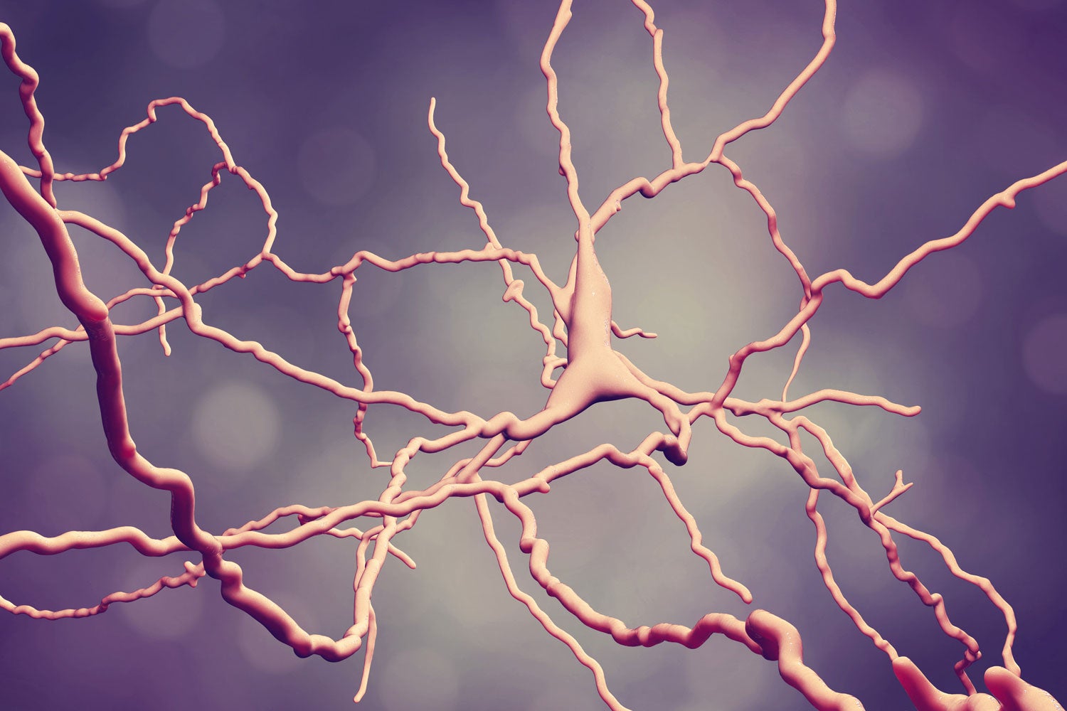
Stanford researchers have found the truth is more complicated than a seminal theory of Parkinson’s disease suggested. (Image credit: Getty Images)
The theory in question, known as the rate hypothesis, has it that Parkinson’s results from an imbalance in brain signals telling the body to start and stop moving.
“The idea was there was too much ‘stop’ and not enough ‘go,’ and that’s why there’s difficulty with movement,” said Mark Schnitzer, the study’s senior author and an associate professor of biology and of applied physics and an investigator of the Howard Hughes Medical Institute.
But that’s only part of the story, said Schnitzer, who co-directs the Stanford Cracking the Neural Code Program and is also a member of Stanford Bio-X and the Stanford Neurosciences Institute. In fact, “start” and “stop” signals are more complex and structured than the rate hypothesis suggests, and Parkinson’s disease in part reflects a loss of that complexity and structure.
New techniques to test an old idea
Doctors have known for decades that Parkinson’s involves the loss of neurons in a region of brain called the substantia nigra, and that the loss affects brain circuits thought to be responsible for initiating and terminating movement. With that in mind, the rate hypothesis seemed quite reasonable: If there was abnormal neural activity in the start or stop circuits, that could lead to the movement problems associated with Parkinson’s.
But testing that hypothesis proved difficult, because the neurons that make up the two pathways are closely intertwined. To see if the start pathway neurons were in fact suppressed, as the rate hypothesis suggested, while stop pathway neurons were overactive, researchers needed a way to track the activity of individual neurons.
To do so, Schnitzer, research associate Jones Parker, Schnitzer’s former graduate student Jesse Marshall and colleagues turned to mice that had been genetically modified so that neurons in the start and stop pathways would flash green when they were active. The team examined the mice under three distinct conditions: normal healthy conditions; a condition that mimics Parkinson’s disease; and that same Parkinson’s-like condition but this time treated with L-dopa (levodopa), the most common drug for Parkinson’s symptoms.
Then, the team peered into their mice’s brains with miniature, head-mounted microscopes to look for green flashes of light that indicate what the start and stop neurons were up to.
Shining light on the details
There were surprises almost immediately. “We found all this undiscovered structure” in both pathways, Marshall said. Rather than all neurons in one or the other pathway lighting up at once, as the rate hypothesis would suggest, certain clusters seemed to be associated with certain activities. In healthy mice, a cluster in the start pathway might light up as a mouse began to turn left, while another in the stop pathway might light up when that mouse finished grooming its tail.
There were more surprises in the mice that mimicked Parkinson’s disease. Although there was less activity in the start pathway, as the rate hypothesis predicted, activity in the stop pathway became unstructured. Rather than suppress particular movements – “stop grooming” or “stop turning left,” say – the stop pathway now seemed to be suppressing many different movements at once.
Treating those mice with L-dopa restored normal activity in both start and stop pathways, the team found, but things went wrong if the dose was too high. Now, there was less activity in the stop circuit, while activity in the start circuit lost its structure, so that now it would initiate movements somewhat at random rather than in the coordinated way characteristic of healthy mice. That finding could help explain one of the most common and visible side effects of the treatment of Parkinson’s disease – jerky, uncontrollable movements known as dyskinesia – Schnitzer said.
Beyond Parkinson’s
The idea that Parkinson’s disease affects not just the level but also the structure of activity in start and stop circuits could change how researchers think about Parkinson’s and a number of other diseases – among them Huntington’s disease, Tourette’s syndrome, chronic pain and even schizophrenia – thought to share a similar underlying mechanism, Parker said.
Beyond understanding those conditions better, the results could ultimately lead to better outcomes for patients with those diseases, Schnitzer said. In particular, additional tests comparing L-dopa to two other, less effective Parkinson’s drugs showed how L-dopa fully restored activity in neurons that control movement, while the others did not – hinting that it may be possible to screen new medications by examining their effects on patterns of brain activity.
“So what we may have here is a new way for testing and screening new drugs by looking directly at neural circuit activity,” Schnitzer said.
Additional Stanford authors include Jun Ding, an assistant professor of neurosurgery and of neurology; postdoctoral fellows Benjamin Grewe, Yu-Wei Wu and Jin Zhong Li; graduate students Biafra Ahanonu and Tony Hyun Kim; and research scientist Yanping Zhang. Michael Ehlers, a senior vice president at Pfizer at the time the study was conducted, is also an author.
Marshall is now a postdoctoral fellow at Harvard University. Grewe is now a professor in the Institute of Neuroinformatics at the University of Zurich. Li is now at Cegedim Bio-Engineering. Ehlers is now executive vice president for research and development at Biogen.
The research was supported by the Howard Hughes Medical Institute, the Stanford Cracking the Neural Code Program, the Stanford Photonics Research Center, Pfizer, a GG Technologies gift fund and fellowships from Stanford, the Helen Hay Whitney Foundation, the U.S. National Institutes of Health, the Howard Hughes Medical Institute and the Swiss National Science Foundation.
Schnitzer is a scientific co-founder of Inscopix Inc., which produces the miniature microscope technology used in the study.
-30-
|