|
March 7, 2013
Stanford psychologists uncover brain-imaging inaccuracies
Traditional methods of fMRI analysis systematically skew which regions of the brain appear to be activating, potentially invalidating hundreds of papers that use the technique. By Max McClure
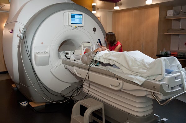
The researchers found that traditional methods of processing fMRI data may lead scientists to overlook smaller brain structures, thus skewing their results. (Photo: Linda A. Cicero / Stanford News Service)
Pictures of brain regions "activating" are by now a familiar accompaniment to any neurological news story. With functional magnetic resonance imaging, or fMRI, you can see specific brain regions light up, standing out against the background like night owls' apartment windows.
It's easy to forget that these brain images aren't real snapshots of brain activity. Instead, each picture is the result of many layers of analysis and interpretation, far removed from raw data.
"It's just one representation of brain activity," said Matthew Sacchet, a PhD student in the Neurosciences Program at the Stanford School of Medicine. "As you process the data, it can change."
Sacchet works in the lab of Stanford psychology Associate Professor Brian Knutson, who studies reward processing in a small area of the brain known as the nucleus accumbens. Precisely how that structure activates is at the heart of an ongoing debate about reward circuits – a subject that holds relevance for our understanding of everything from addiction to financial risk-taking.
Unfortunately, according to a paper from Knutson and Sacchet, hundreds of research papers on this circuit may be unintentionally biased. When the labs processed their fMRI findings, many used a one-size-fits-all strategy that skewed which regions of the brain appeared to be activating.
"I honestly think most people want good data," said Knutson. "I'm excited that we can make this kind of research more rigorous."
The paper appeared in the journal NeuroImage.
Too much smoothing
Functional magnetic resonance imaging measures changes in blood flow in the brain. It's a powerful tool, but the signal fMRI actually detects – the result of the magnetic differences between oxygenated and deoxygenated blood – is noisy.
Researchers need to statistically process the data in order to make the resulting data interpretable. One of the most common approaches is known as "spatial smoothing," which involves averaging the activity of each brain region with that of its neighbors.
But fMRI has only been in use since the mid-1990s. Many of the most common analyses in use today are holdovers from older, lower-resolution types of imaging and seem to have some undesired effects on the finer-grained signals fMRI can provide.
Knutson and Sacchet found that when researchers process fMRI data with a traditional "smoothing kernel" of 8mm, they end up averaging their images over too large an area. Activity in smaller brain structures can then be overlooked, or even shifted to areas that receive more blood flow and where the blood oxygenation level-dependent signal is stronger.
"It might seem strange that a systematic bias like that could bias the whole field," Knutson said. "But if half the people use 8mm and half use 4mm, you might end up with very different results, and it could add up."
Reward structure
These statistical pitfalls are particularly glaring when studying the small, structurally complex nucleus accumbens.
Findings from the Knutson Lab, which has been using the smaller, 4mm smoothing kernel for years, suggest that different parts of the nucleus accumbens have different functions. The forward portion seems to distinguish between positive or negative stimuli, reacting specifically to rewards. Meanwhile, the rear section responds more to the intensity of the motivation.
While some other labs have corroborated this finding, others only found activation in the rear half of the structure.
These contradictory findings now appear to have been skewed. Because the back of the nucleus accumbens is larger and surrounded by more blood-infused gray matter than the front, the smoothing step made it appear as if all the nucleus accumbens' activity originated far to the rear.
A collaborator in Germany already has taken the paper's advice, Sacchet said. "She had a colleague reanalyze her data and found the same thing we found."
Knutson emphasized that the research paper doesn't mean "the methods are bunk." Simply improving the way scientists process signals can enhance their ability to locate specific brain functions.
"There may be a debate, but you can resolve that debate with data," he said.
-30-
|


