|
August 1, 2012
Stanford researchers dive into the mechanical aspects of living cells
A multidisciplinary team of researchers at Stanford measured mechanical tension at the nanoscale to explore how living cells produce and detect force. The research could lead to a better understanding of how tissues and tumors form and grow, and, ultimately, of how complex living organisms organize themselves. By Andrew Myers
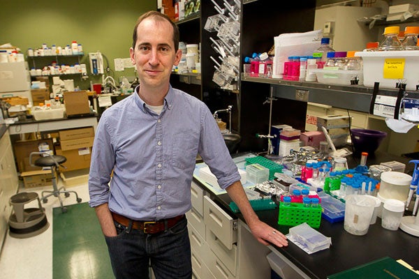
'Cells are really just machines - small, incredibly complex biological machines, but machines nonetheless,' said Alexander Dunn, assistant professor of chemical engineering. (Photo: Norbert von der Groeben / Stanford School of Engineering)
If certain types of living cells are placed on a microscope slide, the cells will inch across the glass, find their neighbors and assemble themselves into a simple, if primitive, tissue. A new study at Stanford University may help explain this phenomenon, and then some, about the mechanical structure and behavior of complex living organisms.
In the paper published in the Proceedings of the National Academy of Sciences, chemical engineering Assistant Professor Alexander Dunn and a multidisciplinary team of researchers in biology, physiology, chemical engineering and mechanical engineering were able to measure – and to literally see – the mechanical forces at play between and within the living cells.
Pulling back the veil on the exact nature of this mechanism could have a bearing on biological understanding ranging from how tissues and tumors form and grow to the creation of entire complex living organisms.
There are scads of data explaining chemical signaling between cells. "And yet, one of the great roadblocks to a complete knowledge of how cells work together to form tissues, organs and, ultimately, us is an understanding of mechanical forces," said Dunn.
Using a new force-sensing technique, Dunn and team have been able to explore how cells connect to one another and how individual cells control their own shape and movement within larger tissues.
Seeing the force
"Cells are really just machines – small, incredibly complex biological machines, but machines nonetheless," said Dunn. "They rely on thousands of moving parts that give the cell shape and control of its destiny."
The mechanical parts are proteins whose exact functions often remain a mystery, but Dunn and team have helped explain the behaviors of a few.
At its most basic level, a cell is like a balloon filled with saltwater, Dunn said. The exterior of the cell, the balloon part, is known as the membrane. Protruding through the membrane, with portions both inside and outside the cell, are certain proteins called cadherins.
Outside of the cell, cadherins bind one cell to its neighbors like Velcro. The "herin" portion of the name, in fact, shares a Latin root with "adhere."
On the inside of the cell, cadherin is connected to long fibers of actin and myosin that stretch from membrane to nucleus to membrane again. Actin and myosin work together as the muscle of the cell, providing tension that gives the cell shape and the ability to control its own movement. Without this force, the balloon of the cell would be a shapeless, immobile blob.
Puppeteer's string
"If you watch a cell moving across a glass slide, you can see it attach itself on one side of the cell and detach on the other, which causes a contraction that allows the cell to, bit by bit, pull itself from place to place," said Dunn. "It's clearly moving itself."
While it was understood that cadherin and actin are connected to one another by other proteins known as catenins, what was not known was how, when and where the cells might be using their muscles (actin and myosin) to tug on the Velcro (cadherin) holding them to other cells.
Dunn and his colleagues have shown for the first time that the actin-catenin-cadherin structure transmits force within the cell and, further, that cadherin can convey mechanical forces from one cell to the next. This is an important problem in the development of organisms, since a cell must somehow control its shape and its attachments to other cells as it grows, divides and migrates from one place to another within the tissue.
Like the strings of a puppeteer controlling a marionette, this mechanism is a form of mechanical communication. Dunn and others in the field believe that these mechanical forces may be important in conveying to a cell how to position itself within a tissue, when to reproduce and when to stop as the tissue reaches its proper size and shape.
"That is the theory, but an important piece was missing," said Dunn. "Our research shows that forces at cell-cell contacts can in fact be communicated from one cell to its neighbors. The theorized mechanical signaling mechanism is feasible."
Story within a story
How Dunn and his colleagues got to this point is a story in itself. It reads like the recipe for a witch's potion – cultured canine kidney cells, DNA from jellyfish and spider silk, and microscopic glass needles.
To measure the force between cells, a team combining the skill of several Stanford laboratories used a tiny and ingenious molecular force sensor developed by Martin Schwartz and colleagues at the University of Virginia. The sensor combines fluorescent proteins from jellyfish with a springy protein from spider silk. The participating Stanford labs are headed by Dunn in chemical engineering, Professors William Weis and W. James Nelson in molecular and cellular physiology, and Associate Professor Beth Pruitt in mechanical engineering.
The genes for the sensor are incorporated into the studied cells' DNA. Under illumination, the cells glow in varying colors depending on how much stretch the sensor is under. In this study, the force sensor is inserted into the cadherin molecules binding cells together – when the Velcro stretches, so does the sensor.
The team then took things a step further. By turning the activity of myosin, actin and catenin on and off, they were able to determine that these proteins are in fact linked together and are at the heart of inter- and intra-cellular mechanical force transmission.
Lastly, using glass microneedles, the team tugged at connected pairs of cells, pulling at one cell to show that force gets communicated to the other through the cadherin interface.
"At this point, we now know that a cell exerts exquisite control over the balance of its internal forces and can detect force exerted from outside by its neighbors, but we still know next to nothing about how," said Dunn. "We are extremely curious to find out more."
Stanford postdoctoral scholar Nicolas Borghi, laboratory technician Maria Sorokina and staff scientist Olga Shcherbakova also participated in the research.
This research was made possible by funding from a Stanford Bio-X Interdisciplinary Initiatives Program award, the National Science Foundation, the National Institutes of Health and a Burroughs-Wellcome Career Award at the Scientific Interface.
Andrew Myers is associate director of communications at the School of Engineering.
-30-
|


